
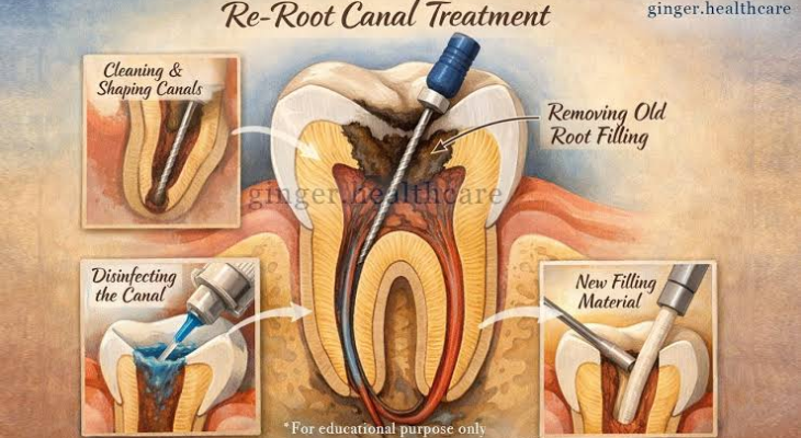
You may have been told you need a root canal, then heard words like curved canals, extra roots, calcified canals, or retreatment. That is usually the moment the worry sets in. You are not just thinking about pain. You are thinking about whether the tooth can be saved, how hard the procedure will be, and whether a general dentist or an endodontist is the right person for the job. If you are considering endodontic treatment in Bolingbrook, these are common and understandable concerns.
That concern makes sense. Teeth do not all follow the same pattern, and the inside of a tooth can be far less simple than the outside suggests. Some teeth have narrow passages, hidden branches, sharp curves, or canals blocked by years of calcification. How endodontists handle complex root canal anatomy comes down to training, imaging, magnification, and technique. The goal is simple. Find the full canal system, clean it well, seal it tightly, and help you keep your natural tooth.
Complex root canal anatomy changes the entire treatment plan
A standard root canal is already precise work. An endodontist is treating a space inside the tooth that can be thinner than a pin. When the anatomy is unusual, the margin for error gets smaller. A missed canal can leave infected tissue behind. A sharply curved canal can make cleaning harder and raise the risk of instrument separation. A calcified canal can be difficult to even locate without the right tools.
This is one reason endodontists focus only on problems inside the tooth. They spend years learning how to manage anatomy that does not follow the textbook version. According to the NCBI overview of root canal treatment, success depends on careful diagnosis, cleaning, shaping, disinfection, and sealing of the canal system. If the anatomy is hidden or unusual, every one of those steps becomes more demanding.
You might hear the term challenging root canal anatomy when a tooth has accessory canals, C shaped canals, severe curvature, open apices, resorption, or prior treatment that altered the original path. Molars are common troublemakers because they often have multiple roots and canals, and some canals are easy to miss without magnification and advanced imaging.
Advanced imaging helps endodontists find canals that standard X rays can miss
Traditional dental X rays show a flat image of a three dimensional structure. That works well for many cases, but not all. Endodontists often use cone beam CT scans when they need a clearer map. A CBCT scan can reveal extra canals, fractures, unusual root shapes, and the true direction of curved roots. That matters before treatment starts and sometimes during retreatment, when old filling material or posts can hide the anatomy.
Research in this published review on root canal anatomy and imaging explains why identifying anatomic variation is central to successful care. If a canal is never found, it cannot be disinfected or sealed. That is often the difference between a tooth that settles down and one that keeps flaring up.
Magnification matters just as much. Endodontists commonly use operating microscopes to locate tiny canal openings, inspect cracks, and work through calcified areas with more control. What looks like a single canal on a regular exam may actually divide into two or more branches deeper in the root.
Specialized tools make difficult root canal treatment safer and more precise
Complex anatomy is not handled by force. It is handled by control. Endodontists use fine ultrasonic tips, flexible nickel titanium files, irrigation systems, and biocompatible filling materials to navigate narrow or curved canals while removing bacteria and tissue debris.
Take a tooth with a sharp curve near the tip of the root. A stiff instrument is more likely to ledge the canal or move off the natural path. A flexible file follows the canal more closely. In a calcified tooth, the issue is access. The canal may be partly closed off, so the endodontist works slowly under magnification to uncover it without weakening the tooth.
This is where endodontic treatment for complex canals differs from routine care. The treatment is not only about reaching the end of the root. It is about preserving the original anatomy as much as possible while disinfecting every reachable part of the system.
Symptoms and treatment difficulty do not always match
Some of the hardest cases are not the most painful ones. A tooth can have very little discomfort and still contain hidden infection, old untreated canals, or a crack that complicates treatment. Other teeth hurt intensely even when the anatomy is fairly direct. That mismatch can be confusing when you are trying to judge urgency by symptoms alone.
The MedlinePlus page on root canal treatment explains the basic reason treatment is needed. The pulp inside the tooth becomes inflamed or infected, often from deep decay, repeated procedures, trauma, or cracks. Once bacteria reach that inner space, the problem does not resolve with home care. If anatomy is complex, delaying treatment can give the infection more time to spread into surrounding bone.
Key differences between routine and complex root canal care
| Factor | Routine Root Canal | Complex Root Canal Case |
| Canal shape | Fairly straight, easier to trace | Curved, branching, narrow, or calcified |
| Imaging needs | Standard dental X rays may be enough | CBCT often helps reveal hidden anatomy |
| Tools used | Standard files and irrigation | Microscope, ultrasonics, flexible files, advanced irrigation |
| Risk of missed anatomy | Lower | Higher without specialist care |
| Typical provider | General dentist or endodontist | Often best managed by an endodontist |
| Time and planning | More straightforward | Greater planning, precision, and follow up |
Clear steps help you move forward with less stress
1. Ask what makes your tooth complex. Get specific words from the exam. Is the issue extra canals, retreatment, calcification, curvature, a post, or a possible crack? When you know the reason, the recommendation makes more sense and the fear becomes easier to manage.
2. Ask whether 3D imaging or specialist referral is appropriate. Not every case needs a CBCT scan or an endodontist, but many difficult molars and retreatments do. If your dentist is already suggesting a referral, that usually reflects the anatomy, not a hopeless tooth.
3. Do not wait for severe pain before acting. A quiet tooth can still be infected. Early treatment often means fewer complications, better odds of saving the tooth, and less chance of swelling or bone loss.
Root canal care gets a bad reputation because people picture pain and uncertainty. The truth is more reassuring. A skilled endodontist is trained for the hidden, curved, blocked, and hard to reach spaces that make some teeth difficult to treat. If you have been told your case is more involved, that does not mean you are out of options. It usually means your tooth needs the right level of precision. Reach out to an endodontist to discuss the anatomy of your tooth, your imaging, and the best next step for saving it.

